Download our TPLO client leaflet here

What is Cruciate Ligament Disease (CCL)?

The cranial cruciate ligament (CCL) is a band of tough fibrous tissue that attaches the front of the femur (thigh bone) to the back of the tibia (shin bone).  It prevents the femur moving backward relative to the tibia and helps to prevent over-rotation (inward) of the knee joint.

What causes CCL?

The most common cause of cranial cruciate ligament disease is a slow degeneration or fraying of the ligament (similar to fraying of a rope).  Other factors may include obesity, individual conformation, hormonal imbalances and inflammatory conditions of the joint.

Which breeds are affected:

In the vast majority of dogs, the ligament ruptures due to degeneration of the fibres within the ligament.  Whilst any dog breed can be affected, there are some breeds which appear to be genetically pre-disposed, we currently do not know the exact reason for this however it is suggested that this is an inherited component related to their gait or conformation.

The breeds at higher risk are:

  • Labradors
  • Rottweilers
  • Boxers
  • West Highland White Terriers
  • Newfoundlands

What are the signs of CCL disease?

Limping (lameness) is the most common sign of CCL disease and it may appear suddenly, have a slow progression or in some cases be intermittent.  Affected dogs may also sit with their leg out to the side.

In some cases it affects both knees at the same time, dogs may find it difficult to rise or stand and can have a ‘pottery gait’.

Unfortunately, if a dog develops it in one knee then it has approximately a 50% chance of developing it in the other knee.

What is happening inside the affected knee?

The fraying of the ligament causes inflammation within the knee and at the time of diagnosis osteoarthritis is often already present.  Alongside the painful lameness caused by the inflammation, a mechanical lameness develops due to the shape of the tibia.  The top of the tibia has a backward slope, when the CCL is intact it helps to keep the femur in the right position on top of the tibia but when the ligament is diseased the femur can slide backward on this slope causing instability within the knee.

How is it diagnosed?

Diagnosis is based on clinical examination by one of our experienced orthopaedic surgeons.  By performing specific manipulations of the knee joint they can establish whether there is laxity.

Radiographs of the affected knee will support this diagnosis and allow the Vets to make a plan for surgery.

Diagnosis can only be 100% confirmed by directly visualising the ligament at the time of surgery.

How can CCL be treated?

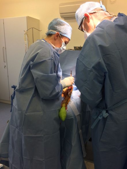
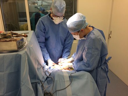
Here at The Meopham Veterinary Hospital, Sevenoaks, Swanscombe & Tonbridge Veterinary Surgeries a Tibial Plateu Leveling Osteotomy (TPLO) procedure is, in most cases, our chosen method for repair.  This is based on medical research showing recovery after TPLO surgery is advantageous over other techniques with a shorter recovery in the short-term and better long-term outcome.

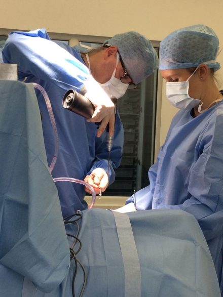
TPLO surgery involves a semi-circular cut in the top of the tibia, the segment is then rotated to change the slope of the top of the tibia.  This prevents the femur ‘sliding’ off the back of the tibia.  The bone is then fixed in place with a plate and screws in the new position.

A crucial part of the surgery is inspecting the inside of the joint where both the ligament and the cartilage are assessed.  If the ligament is completely ruptured the loose ends are removed.  When the joint is unstable the bones start to rub on the cartilage in inappropriate ways and this can cause tearing of the cartilage.  This can be present at the time of surgery or can develop afterwards, which is a common cause of failure to improve or improving then deteriorating after surgery.

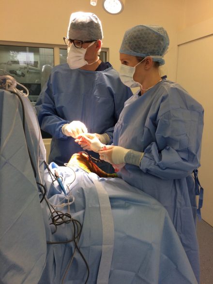
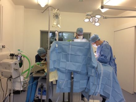
All TPLO procedures are carried out by Dr Martin Hobbs, Dr Penny Barnard-Brown and Dr Rupert Davenport at our Meopham Hospital.  They all continued with post graduate study and have each gained a certificate in small animal surgery.

As a general rule approximately 90% of dogs will return to normal activity after a TPLO.

The surgery is not the only part of treatment, to achieve the best results a strict exercise and rehabilitation regime is also essential.  Full recovery can take 3-6months.

Image taken from lecture notes from Depuy Synthes Course by Michael Kowaleski

Rehabilitation

Here at The Meopham Veterinary Hospital, Sevenoaks, Swanscombe & Tonbridge Veterinary Surgeries we use K-Laser  therapy to aid recovery for all orthopaedic procedures.  Patients will receive therapy immediately post-operatively, the day after surgery before they are discharged home & 3-5 days post-operatively combined with their wound check.  These sessions are carried out by our Registered Veterinary Nurses, further information on this service can be found on our dedicated page https://meophamvets.co.uk/small-animal/laser-therapy/

The first 6 weeks following TPLO procedure require the greatest degree of restriction in the dogs’ activity levels.  Animals should be confined to one room or a cage and taken outside on a lead for toilet purposes only, they must be restricted from activities likely to overload the repair.

Clinical and radiographic examination of your dog is required 6 weeks post operatively.  This will allow clinical assessment and discussion of your dog’s progress and future plans.

In the vast majority of cases animals can usually start to increase their activity levels following this visit and may also benefit from physiotherapy & hydrotherapy at this stage.  The aim is for a full return to exercise 3 to 4 months following surgery.

For further information on our in house Hydrotherapy suite please visit our dedicated page  https://meophamvets.co.uk/hydrotherapy/

What are the potential complications?

All surgeries carry some potential complications.  The two most common complications after surgery are infection and mechanical complications.  Infection is treated by using antibiotics; in some cases surgical irrigation is necessary and in the worst case scenario the implants may need to be removed.

Mechanical complications occur when the dog stresses the plate and screws too much before the bone has healed, which is normally around 6 weeks.  Post-operative radiographs are taken at 6weeks to confirm the healing.  In some cases mechanical complications require surgical revision.

How to book or make an enquiry.

For further information please do not hesitate in contacting us at referrals@meophamvets.co.uk or calling 01474 815333.

We are a family owned, independent RCVS accredited Small Animal Hospital.  We are situated in North Kent and are easily accessible from both the M20 and the A2.  Conveniently located on the A227 only 15 minutes from Gravesend and less than 20minutes from Bluewater.

We regularly accept referrals from all over the South East including Kent, Essex, Surrey, Sussex and the London Boroughs.